
Introduction
Image segmentation is the process of partitioning a certain image into several regions of interest (ROI). In the Biomedical field segmented images can be used for anomaly detection, diagnosing diseases, computer-integrated surgery, treatment planning, studying anatomical structures, and much more. One of the advantages of having automated image segmentation methods is it’s not expensive and requires less work compared to manual segmentation. Automated image segmentation, when built reliably, can provide accurate and consistent results that is helpful for medical professionals.
Biomedical Image Segmentation
Image segmentation tackles different features, it’s not only focused on colors but also in patterns, textures, orientations, and other features. In terms of medical images, there are also different kinds of them produced using different medical imaging acquisition technologies like ultrasonography, x-rays, mammography, computed tomography (CT scans), magnetic resonance imaging (MRI) ,and nuclear medicine.
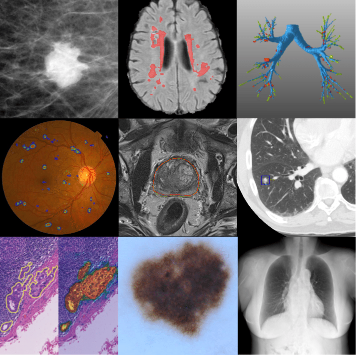
Medical images are also presented in either 2D or 3D domains. 3D medical images differ in sizes and resolution and in some situations they consists off a collection of 2D slices. Image segmentation is a key step in image processing, where all pixels (2D) or voxels (3D) are labeled based on their classification/class or what specific region of interest it is representing. There are different approaches used for image segmentation and based on the complexity of the images on an image’s features like structure, noise, and contrast the performance of the thee different models varies.
Challenges
Some of the challenges that medical image segmentation tackles are partial volume effects, noise, insufficient resolution, structure differentiation due to the existence of noise, and inhomogeneity. There are already key technology advances in the medical imaging field but there are still issues that are hard to solve. Naturally human organs have complex structures because they have different shapes and intensities. Adjacent organ structures are connected and have similar image intensities which can be difficult for the segmentation process. Boundaries on connected organs are blurred which is difficult to segment .
Different Image Segmentation Methods
a few commonly used segmentation methods. We will discuss the following :
- Watershed segmentation
- K-Means Segmentation
- Region growing segmentation
- SVM
- KNN
- CNN
1. Watershed segmentation
The method is based on the idea of watersheds where different regions in an image has intensity that can flow out to other regions in the image; the watershed is the barrier between objects in an image from which the intensity "flows" out in all directions different regions.
Watershed image segmentation is a good approach for images with overlapping or touching objects. This is often the case in medical images, where organs or tissues can touch or overlap, making it difficult to separate them using other methods.
However, this method tend to be sensitive to noise and can do over-segmentation thus it is sometimes implemented with other pre-processing techniques. In one study by Liang, Y and Fu, J [1] they proposed an improved watershed segmentation algorithm and performed it on brain lesion MRIs. Their model improved the watershed algorithm’s issues on over-segmentation and noise contamination of images and got good segmentation results on their dataset.
2. K-Means Segmentation
This method partitions an image into K number of clusters, where each cluster is represented by a centroid which represents the center of a cluster. The process first starts with an initial location of the centroids then iteratively updates the centroid locations and the assignment of pixels to clusters until a convergence criterion is met.
In one study by Sammouda and El-Zaart , they implemented K-Means Clustering Algorithm with Elbow Method to segment prostate images to help detect prostate cancer [2] . Their paper aims to segment and analyze pixels of histological and near-infrared (NIR) prostate cancer images. Using k-means clustering algorithm with elbow method their study gave better clustering of pixels through automatically determining the best number of clusters in an image.
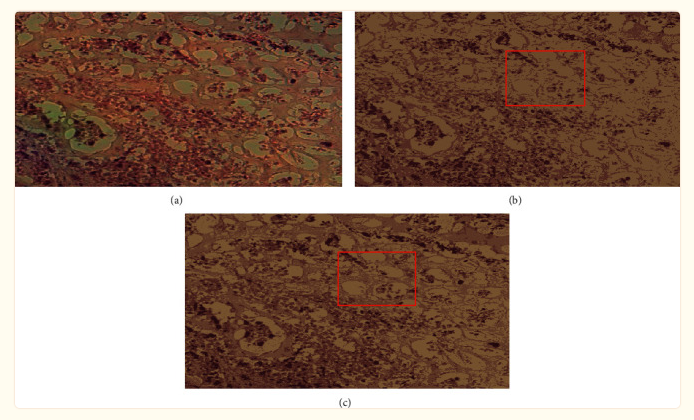 A sample segmented image from Sammouda and El-Zaart's paper. (a) the original image, (b) its segmented image with K = 3, and (c) its segmented image with K = 4.
3. Region growing
This method is based on the idea that pixels in an image have common characteristics with its neighboring pixels.The process will begin with a seed point in an image, then there will be an iterative process where each pixel will be compared to its neighbors and if they have similarities they will be grouped to a similar cluster. The process will continue until it reaches a stopping criterion like reaching the boundary of the image or the maximum size and intensity range for the region.
In one study by Biratu, E.S. ,Schwenker, F., Debelee, et. al, they implemented an enhanced region growing method for brain tumor MR image segmentation [3]. Their proposed method used a thresholding pre-processing technique to seperate the skull from the brain image, afterwards they chose a seed point for the region growing algorithm and obtained five different regions of interest (ROIs) for each skull stripped input brain image. Their approach showed good segmentation results.
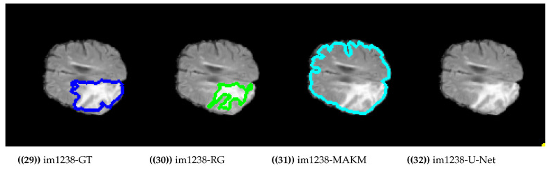 Sample results of their approach in comparison with other algorithms such as MAKM, U-Net, and the image ground truth.
4. Convolutional Neural Networks (CNN)
CNNs is a deep learning algorithm that assigns different objects or instances in an input image to be able to differentiate them from one another. They work by using multiple layers of artificial neurons to learn hierarchical representations of image data.
Deep neural models CNNs shows significant growth in their use for medical image segmentation because of its high performance and accuracy. There is also a gaining popularity on the use of 3D medical image analysis. In one paper by Niyas, et al. [4], they presented a survey on medical image segmentation that uses 3D convolutional neural networks. they dicussed different applications of CNN in segmentation of medical images such as CT scans (lungs, liver, thoracic, and abnomen) , MRI (brain, heart, and and), and Ultrasounds (breast)
5. Support Vector Machines (SVM)
SVM is used to predict the class label of each pixel in the image, where the class labels correspond to different regions or objects in the image. The algorithm works by finding the hyperplane that separates the pixels into different classes, based on their features and intensity values. One of its benefits are it can have significant accuracy with less computation power.
For using SVM for image segmentation, it will segment different regions of interest by predicting the class label for each pixel in the image, and the resulting image can be visualized as a segmented image.
In one study by Khairandish,Sharma, Jain ,et al., they used a hybrid model of CNN and SVM models to classify Benign and Malignant tumors from brain MRI images [5]. They compared accuracy results other models like Deep CNN (DCNN), Deep Neural Network (DNN) and Discrete Wavelet Autoencoder (DWA), k-nearest neighbors (kNN),and CNN. Their hybrid model got the highest accuracy result of 98.49%
References:
[1]Liang, Y., & Fu, J. (2018). Watershed Algorithm for Medical Image Segmentation Based on Morphology and total Variation Model. International Journal of Pattern Recognition and Artificial Intelligence. doi:10.1142/s0218001419540193
[2] Sammouda R, El-Zaart A. An Optimized Approach for Prostate Image Segmentation Using K-Means Clustering Algorithm with Elbow Method. Comput Intell Neurosci. 2021 Nov 15;2021:4553832. doi: 10.1155/2021/4553832. PMID: 34819951; PMCID: PMC8608531.
[3] Biratu ES, Schwenker F, Debelee TG, Kebede SR, Negera WG, Molla HT. Enhanced Region Growing for Brain Tumor MR Image Segmentation. J Imaging. 2021 Feb 1;7(2):22. doi: 10.3390/jimaging7020022. PMID: 34460621; PMCID: PMC8321280.
[4]Niyas, S., Pawan, S. J., Kumar, M. A., & Rajan, J. (2021). Medical image segmentation using 3D convolutional neural networks: A review. arXiv preprint arXiv:2108.08467.
[5]Khairandish, M. O., Sharma, M., Jain, V., Chatterjee, J. M., & Jhanjhi, N. Z. (2021). A Hybrid CNN-SVM Threshold Segmentation Approach for Tumor Detection and Classification of MRI Brain Images. IRBM. doi:10.1016/j.irbm.2021.06.003